- Статьи
- Science and technology
- In the interface area: the digital platform will detect malignant kidney tumors
In the interface area: the digital platform will detect malignant kidney tumors
The Russian service for oncourologists, which helps to make an accurate diagnosis to patients with kidney diseases, has passed the stage of technical tests and will soon be registered for clinical use in the Russian Federation. The web platform automatically combines the four phases of a CT scan into a single three-dimensional image in just a few minutes. There are also plans to train a digital biopsy virtual assistant, meaning he will be able to determine whether a tumor is benign or malignant. For more information about the prospects of the technology, see the Izvestia article.
A service for searching for kidney diseases
A web-based medical decision support platform Sechenov.AI_nephro, which helps to accurately diagnose patients with kidney diseases, has successfully passed technical tests before registration, told Izvestia at Sechenov University. According to doctors, kidney tumor is one of the most common diseases that oncourologists face. About 20 years ago, a patient with this disease had no choice but to have a kidney removed. But gradually, resection, an operation to remove a tumor while preserving an organ, began to enter clinical practice.
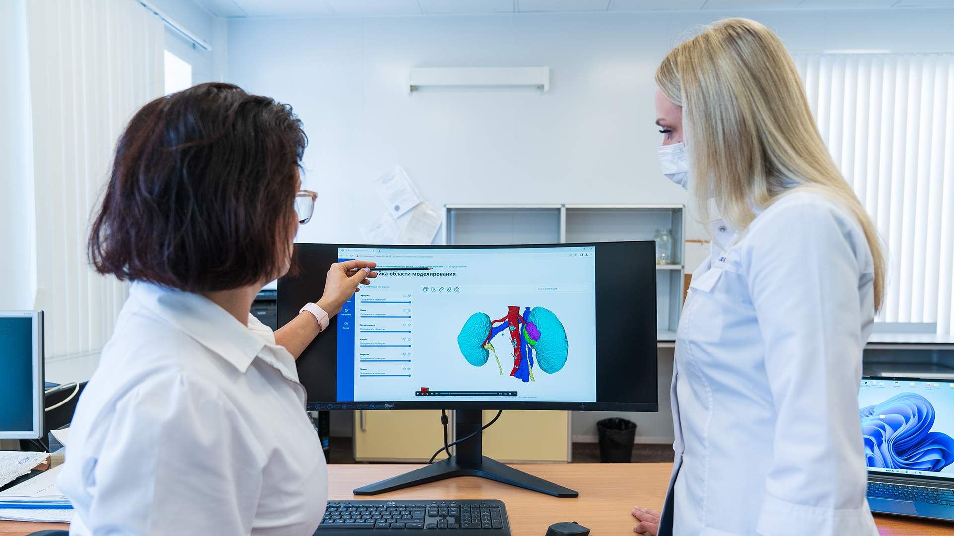
To reduce the risk of complications after such surgery, the surgeon must know the exact location of the tumor and which anatomical structures it borders on. It became clear that a 3D image was required, which would show not only the kidney tumor, but also the vessels that feed it, elements of the pelvic system, veins and neighboring structures. Since 2008, the Institute of Urology at Sechenov University has started using 3D modeling of the pathological process in patients with neoplasms of the parenchyma (tissue of the internal environment) of the kidney. In manual mode, when a team of three specialists — a urologist, a radiologist, and an IT specialist - worked , the construction process took up to a day.

Now, thanks to the development of a new web resource and the automation of the system, obtaining an image is a matter of a few minutes, the university said. The doctor needs to download the CT scan of the patient and select the area of interest in the interface — tumors in the kidney area.
— Before getting to the server, this zone is cut out and sent for processing," a leading software engineer at the University Center for Neural Network Technologies at the Institute of Urology told Izvestia.Ivan Chernenky. — And already on the server side, the neural network identifies the necessary anatomical structures: arteries, veins, ureters, kidney parenchyma, tumors, cysts and creates an additional file with these "masks". After that, it is returned to the user and a 3D construction is already underway that can be worked with.

According to the specialist, thanks to the 3D model, it is possible to estimate the depth of the tumor's immersion in the kidney, perform virtual resection with different planes and obtain other information important to the doctor.
What is a digital biopsy
In the near future Sechenov.AI_nephro will be registered by Roszdravnadzor, the university said. The following options are currently available on the web platform: 3D modeling of the pathological process in patients with renal parenchyma neoplasm, and the system can also monitor patients with kidney cysts and hydronephrosis.
— The plans include training the digital biopsy web platform, meaning it will be able to determine whether a patient has a benign or malignant neoplasm, as well as digital puncture to recognize the type of tumor. By the end of this year, we hope to implement this in the form of an alpha version of the program," Evgeny Sirota, director of the Center for Neural Network Technologies, told Izvestia.

The web platform can also be useful for transplant doctors who are engaged in kidney transplantation. An agreement on cooperation in this field has already been signed with Brest Clinical Hospital (Belarus), the specialist added.
— Colleagues from Brest have accumulated considerable experience in performing kidney transplantation. The share of disciplines that we plan to add to the platform is very important for transplant doctors, both at the planning stage in terms of functional status and at the postoperative stage to assess the functional state of the transplant," he explained.

Currently, the introduction of new high-tech diagnostic methods, such as 3D technology, allows for a more detailed understanding of the individual topographic and anatomical features of the organ structure in combination with the pathological process and course of the disease, said Aldar Ochirov, a urologist at the Semeynaya clinic.
— According to scientists, thanks to the creation of a web platform, the neural network creates within a few minutes a 3D model of the affected area of interest and makes it possible to perform a virtual resection and obtain important information for the surgeon. Indeed, thanks to the additional 3D diagnostic method , a preliminary assessment of kidney cancer surgery allows you to choose the method and access of intervention, plan pre-operative treatment, prevent the development of complications and predict the result, which is of great benefit to both the doctor and the patient ," he noted.p>

Also, in addition to kidney neoplasms, this method allows to identify individual features and structure of the organ, to identify pathologies such as kidney stones, hydronephrosis, cysts and so on, the urologist noted.

Similar solutions exist abroad, such as Virtual Surgical Planning or Mimics Innovation Suite, however Sechenov.AI_nephro stands out for its user-friendly web format, which makes access to the technology as simple and efficient as possible, commented the co-founder of biotech startup studio Scanderm (market participant NTI "HealthNet"). Evgeny Sobolev. A logical step in the development of the platform will be the introduction of algorithms for the diagnosis of bladder cancer.
— This type of cancer is difficult to detect in the early stages: the tumor hides in the mucous membrane and often changes its morphology. In addition, endoscopy videos are saturated with visual noise, which complicates diagnosis even for experienced specialists. If we create high—quality segmentation and markup of data, it will allow us to assemble a powerful training dataset for AI that can automatically detect tumor changes," the expert said.
The project is implemented within the framework of the strategic academic leadership program "Priority 2030" and the strategic project "Network for the Development of Best Practices in Medicine, Science and Education".
Переведено сервисом «Яндекс Переводчик»
