- Статьи
- Science and technology
- Get hooked on spheroids: 3D printed tumor could help find a cure for cancer
Get hooked on spheroids: 3D printed tumor could help find a cure for cancer
Russian scientists have developed a technology for creating realistic models of cancerous tumors - they are printed on a 3D printer. These tissue-engineered constructs are intended for testing drugs and new methods of cancer therapy. Now such experiments are conducted on single-layer cell models. However, due to their two-dimensional structure, they cannot replicate the structure of real tumor tissue. The proposed technique will make it possible to understand how a potential drug substance penetrates deep into the pathological formation. According to experts, the technology is promising, but further complication of the model is needed to achieve breakthrough results.
Realistic tumor model
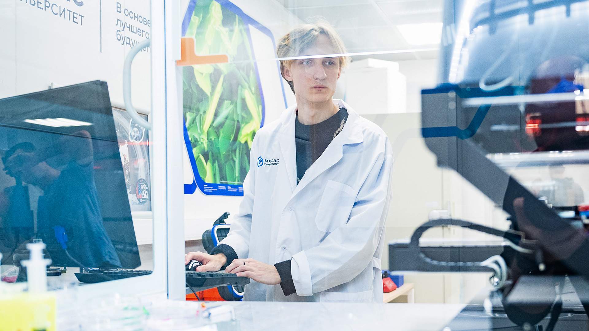
Specialists of NITU MISIS have developed equivalents of cancer tumors using 3D bioprinting. They are intended for detailed study of mechanisms of malignant neoplasms appearance and creation of more effective methods of their treatment. Now in laboratory experiments for these purposes, scientists use models consisting of a monolayer of cells. However, because of their two-dimensional structure, they cannot accurately reproduce the structure of a three-dimensional tumor. Therefore, they cannot trace the efficiency of drug penetration into the depth of the tumor.

- Three-dimensional equivalents of tumor tissue, which could simulate its structure in vitro, are not yet used by pharmaceutical companies. But their creation and introduction into the process of developing new antitumor drugs is just a matter of time," said Elizaveta Kudan, head of the scientific and educational laboratory of tissue engineering and regenerative medicine at the university.
3D models for drug testing are used to simulate human biology in more detail. They can be used to test drug toxicity and create disease scenarios. Such tissue-engineered structures can be developed individually for the anatomy of a particular patient. The use of 3D models will help reduce the need for animal testing and speed up drug development.
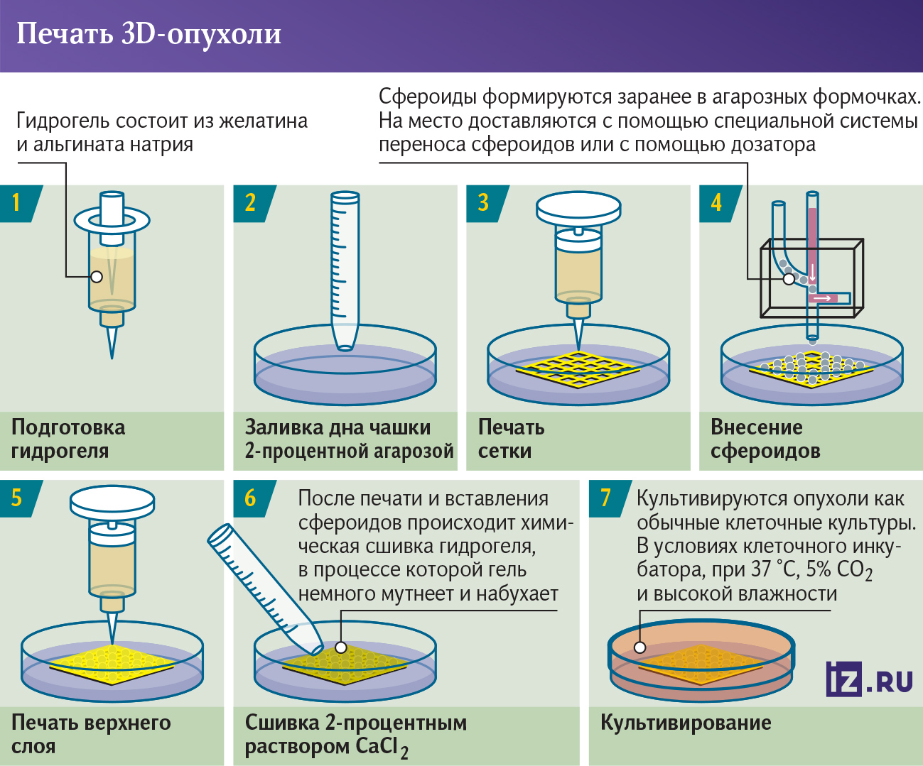
To print the model on a 3D bioprinter, scientists used pancreatic cancer cells and skin cells - fibroblasts. They became the main component of the microenvironment of the malignant neoplasm. The obtained samples remained viable for three to four weeks.
Further complication of the model
The experts also investigated how the properties of the final tissue-engineered structures depend on their structure. The shape affects the tumor microenvironment and cancer progression. In most similar studies, malignant cells are placed in the center and the other components are placed on the periphery. However, this arrangement produces a capsule rather than a complete stromal structure (the backbone of the organ).

- The models were printed using tissue spheroids (tissue-like multicellular aggregates of spherical shape. - "Izvestia"). This is a more complex approach than traditional extrusion bioprinting (one of the most common bioprinting methods, in which bioinks are applied to the substrate using a nozzle). However, the use of full tissue spheroids as miniature building blocks allows us to achieve higher cell densities comparable to native tissues and reduce the time to "mature" the constructs. We are the first to analyze the influence of design and mutual arrangement of cell components on the architecture of models after their maturation, - said Maxim Lugovoi, engineer of the scientific project of the research and educational laboratory of tissue engineering and regenerative medicine at NITU MISIS.

Further work will help scientists to create more realistic models for screening antitumor activity of various substances. In the future, experts plan to complicate the solution - to add vascular systems and immune cells.
- The technology looks interesting: recently, the mechanisms of action of drugs are becoming more and more complex, and it is increasingly difficult to select models for their experimental study. At the same time, models for preclinical studies should be as technically simple, inexpensive and easily reproducible as possible. And printing with the help of tissue spheroids is quite complicated and can be difficult to implement in the routine practice of creating new drugs," said Vadim Pokrovsky, Head of the Department of Biochemistry named after Academician T.T. Berezov of the Russian Academy of Medical Sciences.

Stanislav Ottavnov, Head of the Laboratory for Analyzing Health Indicators at MIPT, agrees that the technology is promising. However, he sees nothing revolutionary in it.
- In fact, scientists have only modified it in some ways, made it more complicated, and made it a little "closer" to life. Such improvement of models is inevitable, and their development is only a matter of time. Intuitively, it seems that this work will accelerate preclinical research and reduce the need for animal studies, but nothing supernova scientists have not offered, just another step on a long road, - he said.
When the model becomes more complex and has different cells, primarily vascular cells, it will be possible to already talk about it as a significant achievement, the expert added.
Переведено сервисом «Яндекс Переводчик»
